Respiratory Examination
The respiratory examination involves an assessment of how a patient's lungs are functioning. Like most examination, it is a systemic examination that looks for signs of respiratory compromise such as breathlessness, chest pain, haemoptysis or wheeze. It is one of the key clinical examinations of OSCE finals. Therefore it's important to have a slick and thorough approach. It is only once your are confident in performing this examination that you can look out for and identify signs of respiratory diseases such as chronic obstructive pulmonary disease (COPD), pulmonary fibrosis, asthma, pneumonia etc.
Introduction
- Wash hands
Wash your hands using the Ayliffe technique
2. Introduce yourself
Introduce yourself and give your name and grade
3. Check patient details
Clarify patient's identity by confirming name and asking for their DOB
4. Describe examination
Explain what examination you are performing and what it involves
5. Gain verbal consent
6. Offer a chaperone
Ask if they would like a chaperone
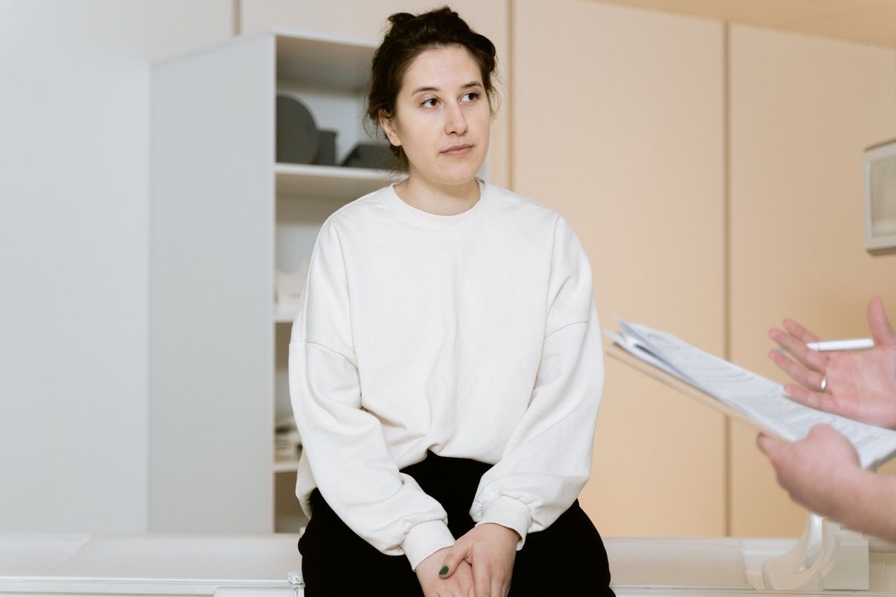
Peripheral Examination
- Positioning patient at 45 deg
Initially lye the patient at 45 degrees and expose them from waist up
2. End of bed inspection
Inspect the patient from the end of the best and look for the following:
Patient
- Breathing (tachypnoea, pattern of breathing, accessory muscles, nasal flaring, subcostal/intercostal recession)Cyanosis (central or peripheral)
- weight loss (cachexia)
- Chest shape (pectus excavatum/carinatum, barrel chest)
Adjuncts
- eg. any supplemental O2 (%), IV lines, infusions, catheter
Paraphernalia
- inhalers, nebulisers, sputum pots, walking aids
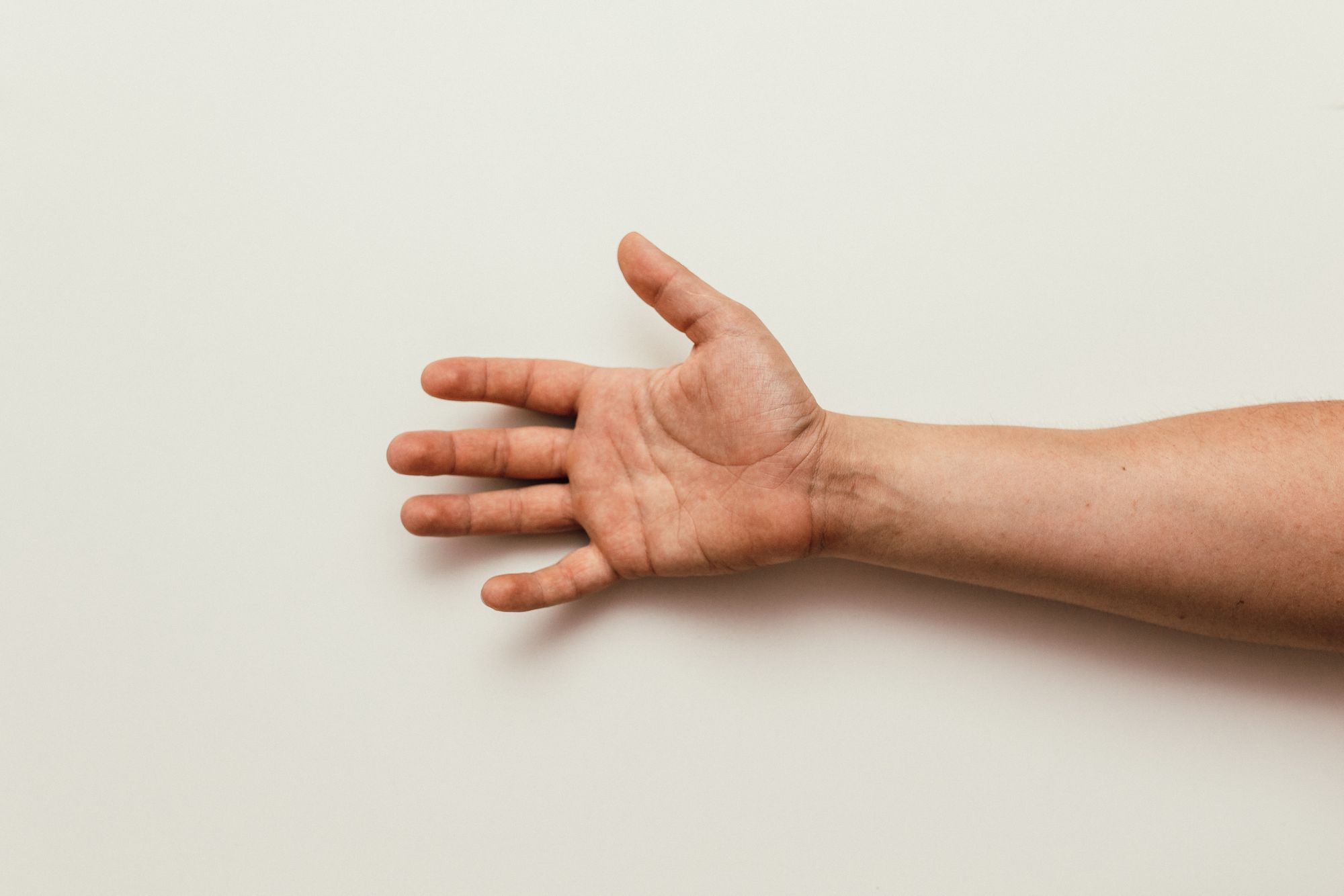
3. Inspect the hands
Inspect the hands and check for stigmata of chronic Respiratory disease
Skin
- Peripheral cyanosis (Poor oxygenation)
- Tar staining (Smoker)
Temperature
- Cold hands - poor circulation (cor pulmonale)
Nails
- Clubbing - Tumours (lung cancer, mesothelioma, pleural fibroma), interstitial lung disease (IPF), bronchiectasis, empyema, lung abscess, CF
- Yellow nails - Yellow nail syndrome, tar staining
- Koilonychia - iron deficiency
4. Check pulse and respiratory rate
Check the patient's pulse and resp rate. Time for 15 seconds and multiply by 4.
- Tachycardia/tachypnoea
- Irregular (AF)
5. Check for tremor and CO2 flap
Ask the patients to put their hands in front of them and check for tremor. Then ask them to cock their wrists back as if they were stopping traffic. Check for any flapping.
- Asterixis (hypercapnia)
- Tremor (excess use of beta-agonist or theophylline)
6. Inspect the face and neck
Next, inspect their eyes, mouth and neck for the following.
Eyes
- Pale conjunctiva (anaemia)
- Ptosis/miosis - Horners syndrome (Pancoast’s Tumour)
Mouth
- Blue tongue (central cyanosis)
- Oral candida (Steroid use)
- Glositis - large tongue (iron deficiency)
- Angular cheilitis - cuts to edge of lips (iron deficiency)
Neck
Raised JVP
- Sign of fluid overload
- Increased intrathoracic pressure - tension pneumothorax
- SVC obstruction - raised and non waveform
Tracheal deviation
- Towards lesion (lobe/lung collapse, fibrosis, pneumonectomy)
- Away from lesion (tension pneumothorax, massive pleural effusion)
Cricoid-sternal space (reduced in hyperinflation eg. COPD)
Lymphadenopathy (Pancoast’s Tumour, Lung Ca, Lymphoma)
Chest examination (FRONT)
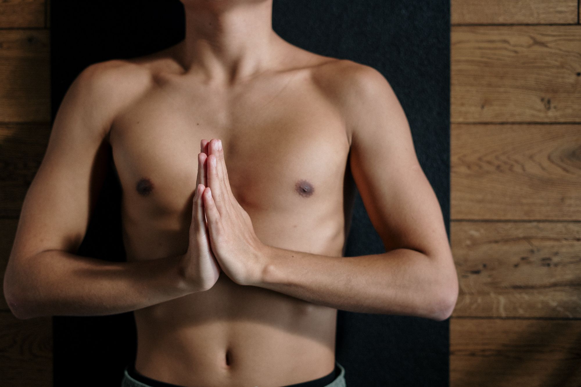
1. General Inspection at front
Inspect the chest again more closely and look for the following:
Breathing
- Use of accessory muscle
- Pattern of breathing
- Paradoxical abdominal breathing - inward movement of abdomen in inspiration (severe COPD)
Shape
- Hyperinflation or barrel chested, pectus excavatum/carinatum
Scar (Pneumonectomy scar, lobectomy, mastectomy)
2. Palpation of the chest at front
Palpate the chest for the following
Apex beat (Normally 5th IC space mid-clavicular line)
- Displaced laterally (enlarged heart ?cor pulmonale)
Chest expansion
- bilateral reduction - COPD and diffuse pulmonary fibrosis,
- unilateral - effusion, collapse, pneumothorax
3. Percussion of the chest at front
Percuss the chest from the front. Make sure you percuss 6 places at the front and 2 places on the side on both the right and left side, comparing as you go along. Go down as far as the 6th rib.
- Hyper resonant - pneumothorax
- Dull - consolidation, collapse,
- Stoney dull - effusion, haemothorax
4. Auscultation of the chest at front
Now listen to the front of the chest over the places you have just percussed. Ask the patient to take deep breaths in and out of their mouth.
- Wheeze - narrowing of distal airways (COPD, Asthma)
- Crackles - peripheral airway collapse (Early inspiratory - Bronchiolitis, Mid inspiratory - pulmonary oedema, Late inspiratory - pulmonary fibrosis, Pan Insp & Exp - Bronchiectasis)
- Bronchial breathing - consolidation around airway (pneumonia, fibrosis)
- Pleural rub - friction between parietal and visceral pleura (pneumonia, pulmonary infarction)
5. Vocal resonance and tactile fremitus at front
Then check for:
- Vocal resonance (ask the patient to say “99” and listen with a stethoscope)
- Tactile fremitus (ask the patient to say “99” and feel the chest wall).
Chest examination (BACK)
Now examine the the patient at the back.
6. General Inspection at back
Inspect the chest again more closely and look for the following:
Shape
- Kyphosis, scoliosis
Scar (Pneumonectomy scar, lobectomy)
7. Palpation of the chest at back
Palpate the chest for the following:
Chest expansion
- bilateral reduction - COPD and diffuse pulmonary fibrosis,
- unilateral - effusion, collapse, pneumothorax
8. Percussion of the chest at back
Percuss the chest wall from the back. Make sure you percuss 6 places on both the right and left side going down as far as the 11th rib.
- Hyper resonant - pneumothorax
- Dull - consolidation, collapse,
- Stoney dull - effusion, haemothorax
9. Auscultation of the chest at back
Now listen to the back of the chest. Make sure you compare the right side to the left. Listen over the 6 places you percussed on both the right and left side.
- Wheeze - narrowing of distal airways (COPD, Asthma)
- Crackles - peripheral airway collapse (Early inspiratory - Bronchiolitis, Mid inspiratory - pulmonary oedema, Late inspiratory - pulmonary fibrosis, Pan Insp & Exp - Bronchiectasis)
- Bronchial breathing - consolidation around airway (pneumonia, fibrosis)
- Pleural rub - friction between parietal and visceral pleura (pneumonia, pulmonary infarction)
10. Vocal resonance and tactile fremitus at back
Then check for:
- Vocal resonance (ask the patient to say “99” and listen with a stethoscope)
- Tactile fremitus (ask the patient to say “99” and feel the chest wall).
End of examination
- Thank patient
Let the patient know you have finished examining them and thank them for their time. Be courteous and offer them help to get redressed.
2. State other exams for completion
Turn to the examiner and state what else you would do to complete the exam.
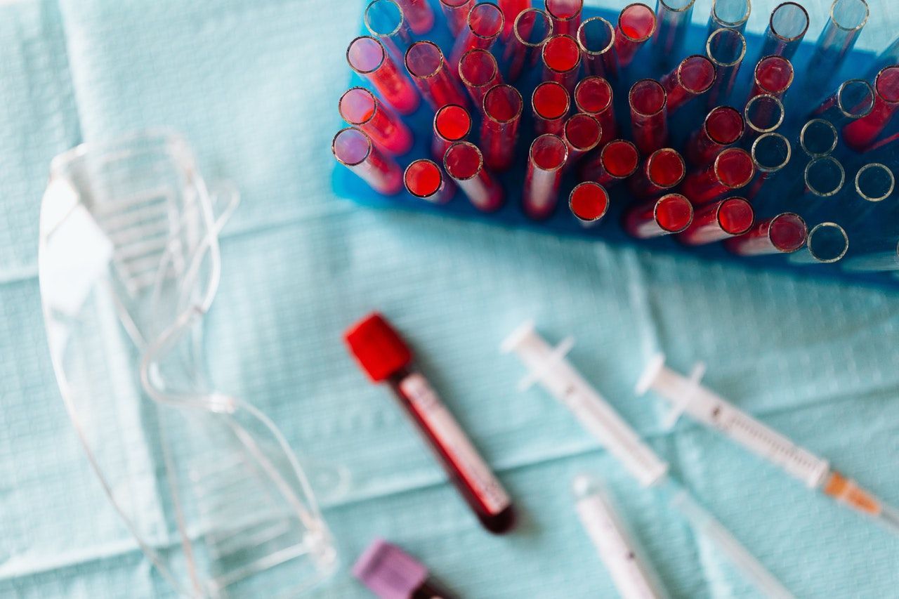
3. State what tests you would perform
Explain to the examiner what tests and investigations you would perform
- Dip - Proteinuria
- Urine - Atypical pneumonia screen
- MC&S
- FBC - anaemia/infection
- U&E - baseline
- LFT - Signs of sepsis, signs of metastases
- CRP / ESR - signs of inflammation
- Atypical pneumonia screen
- Pneumothorax, signs of consolidation/effusion/collapse, metastases. Cardiomegaly / pulmonary oedema.
Doctor Khalid Newsletter
Join the newsletter to receive the latest updates in your inbox.
