Cranial Nerves Examination
The cranial nerve examination is part of the neurological exam. It is useful in identifying pathology in the 12 pairs of cranial nerves that come from the brain. The ability to test them in a systematic and slick manner is one of the key requirements as a medical student. Although often neurological examinations are dubbed as the harder ones in OSCEs, but if you begin by understanding what each nerve does and what signs/symptoms to look for then it becomes a case of pattern recognition. Pathology found in cranial nerve examinations can vary from things like Multiple Sclerosis (MS), trigeminal neuralgia or space occupying lesions.
Introduction
- Wash hands
Wash your hands using the Ayliffe technique
2. Introduce yourself
Introduce yourself and give your name and grade
3. Check patient details
Clarify patients identity by confirming name and asking for their DOB
4. Describe examination
Explain what examination you are performing and what it involves
5. Gain verbal consent
Peripheral Examination
- Positioning patient at 45 deg or sit them up
Lye the patient at 45 degrees or sit them up
2. End of bed inspection
Inspect the patient from the end of the best and look for the following:
Patient:
- Facial symmetry - any droop, protruding tongue
- Speech - Are they understand you? Can they communicate verbally?
- Eyes - are they able to close both lids? Are the eyes watering? Are the pupils the same size?
Paraphernalia:
- glasses, walking aids
Cranial Nerve Examination
1. Cranial Nerve I - Olfactory Nerve
This is not always tested in an exam setting. However if you are presented with smelling salts the test should be undertaken.
Ask the patient to close their eyes and place common odours under their nose e.g. orange peel, chocolate, coffee, lemon peel, and ask them to identify the scents.
Anosmia:
- Loss of the sense of smell
- Head injury
- Skull base tumours
- Parkinson’s Disease
- Huntington’s disease
Parosmia:
- Altered sense of smell
- Head trauma
- Sinus infection
- Iatrogenic causes
- Temporal lobe epilepsy
2. Cranial Nerve II - Optic Nerve
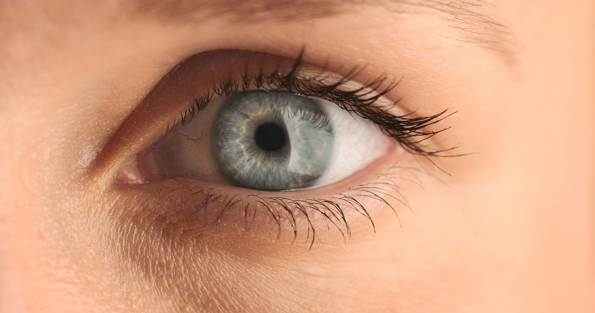
Next move on to look at the eyes and test the optic nerves. Here you will be testing for the following:
General inspection
Conduct a general inspection of the eyes looks for the following.
Eyelids:
- Any ptosis (unilateral - Horner’s syndrome, bilateral - Argyll-Robertson pupils)
- Exophthalmos (Thyrotoxicosis)
Pupils:
- Unilateral miosis - constricted pupils (Horner’s syndrome)
- Bilateral miosis (Argyll-Robertson pupils)
- Unilateral mydriasis - dilated pupils (Holmes-Adie pupil, SOL, head injury, orbital trauma)
- Bilateral mydriasis (iatrogenic - anticholinergics, cocaine)
Position:
- “Down and out” (Third Nerve palsy)
Pupillary reflex to light
Next shine a light into the one of the pupils and see if it constricts. Shine a light again onto the same side, but this time checking if the opposite pupil constricts. Repeat the steps again on the opposite eye.
- (R) optic nerve injury - Light shone in (R) eye (neither pupils constrict), light shone in (L) eye (both pupils constrict)
- (R) oculomotor nerve injury - When a light is shone in either eye, only the (L) pupil constricts (i.e. (R) pupil can’t constrict)
- (L) optic nerve injury - Light shone in (L) eye (neither pupils constrict), light shone in (R) eye (both pupils constrict)
- (L) oculomotor nerve injury - When a light is shone in either eye, only the (R) pupil constricts (i.e. (L) pupil can’t constrict)
Accommodation
Check the pupils ability to converge and focus from a distant object to an object that is closer to the face. Ask the patient to look at the wall, then place a finger in front of the patient and ask them to focus on it. The eyes should converge (come together) and the pupils should focus by constricting.
- Convergence (Oculomotor nerve)
- Focus (Optic nerve)
Visual acuity
Test the patients vision using a standardized Snellen Chart. The patient’s vision should be tested both with their visual aids and without. Document their score.
Visual fields
Illicit the patient's visual fields by asking them to close one eye and to focus on one of yours. Test each quadrant of the visual field by bringing in a moving finger and asking the patient when they are able to visualize it. Document their field of vision.
For completion fundoscopy should be completed at this point. In an Exam setting this is not usually requested. However, do so if provided with an ophthalmoscope.
3. Cranial Nerve III, IV, VI - Oculomotor Nerve, Trochlear Nerve, Abducens Nerve
Some of the functions of the Oculomotor Nerve have already been tested. The motor function of the Oculomotor Nerve can ve tested at the same time as the Trochlear and Abducens Nerve.
Ask the patient to focus on your finger and to keep their head still. Then ask them to follow your finger as you draw 2 large “H” shapes in front of them. Ask if they experience any double vision and note any nystagmus.
- (R) Oculomotor Nerve injury - (R) eye will be down and out, unable to look up
- (R) Trochlea Nerve injury - Inability to look down and in with (R) eye. Will get double vision looking down and medially.
- (R) Abducens nerve injury - inability to abduct the (R) eye. Therefore patient will get double vision gazing laterally.
The same will occur on the (L) eye.
Cranial Nerve V - Trigeminal Nerve
Next move onto examining the Trigeminal Nerve which has three branches (Opthalmic branch, Maxillary branch and Mandibular branch). They provide sensation to the face and the motor supply for the muscles of mastication. The Trigeminal Nerve also provides the reflex pathway for the corneal and jaw reflex.
Sensation:
- Examine the three branches over the jawline (Mandibular branch), cheek line (Maxillary branch) and forehead (Ophthalmic branch) for fine touch and pain.
Assess pain using a Neurotip if provided. If not test light and course touch.
Motor:
- Test the muscle of mastication by asking the patient to clench their teeth. Assess muscle bulk and tone of the temporalis muscle and buccinator. Compare right to left.
Reflex:
- Corneal reflex - Dab the eyeball with cotton wool. Negative finding if eyelid’s don’t close (Won’t if there is damage to Ophthalmic branch)
- Jaw jerk - place a finger on the lower jaw and strike with a tendon hammer. Positive if jaw clenches for a prolonged time (indicates an upper motor neurone lesion)
5. Cranial Nerve VII - Facial Nerve
Move on to testing the Facial Nerve which provides the motor branches of the facial muscles for expression.
Ask the patient do the following movements and then resist against opposition:
- Raise eyebrows
- Close eyes
- Blow out their cheeks
- Smile and show their teeth
If there is weakness on one side, deduce whether it is due to an upper or lower motor neurone lesion.
- Upper motor neurone lesion - sparing of forehead muscles on affected side
- Lower motor neurone lesion - forehead not spared
6. Cranial Nerve VIII - Vestibulocochlear Nerve
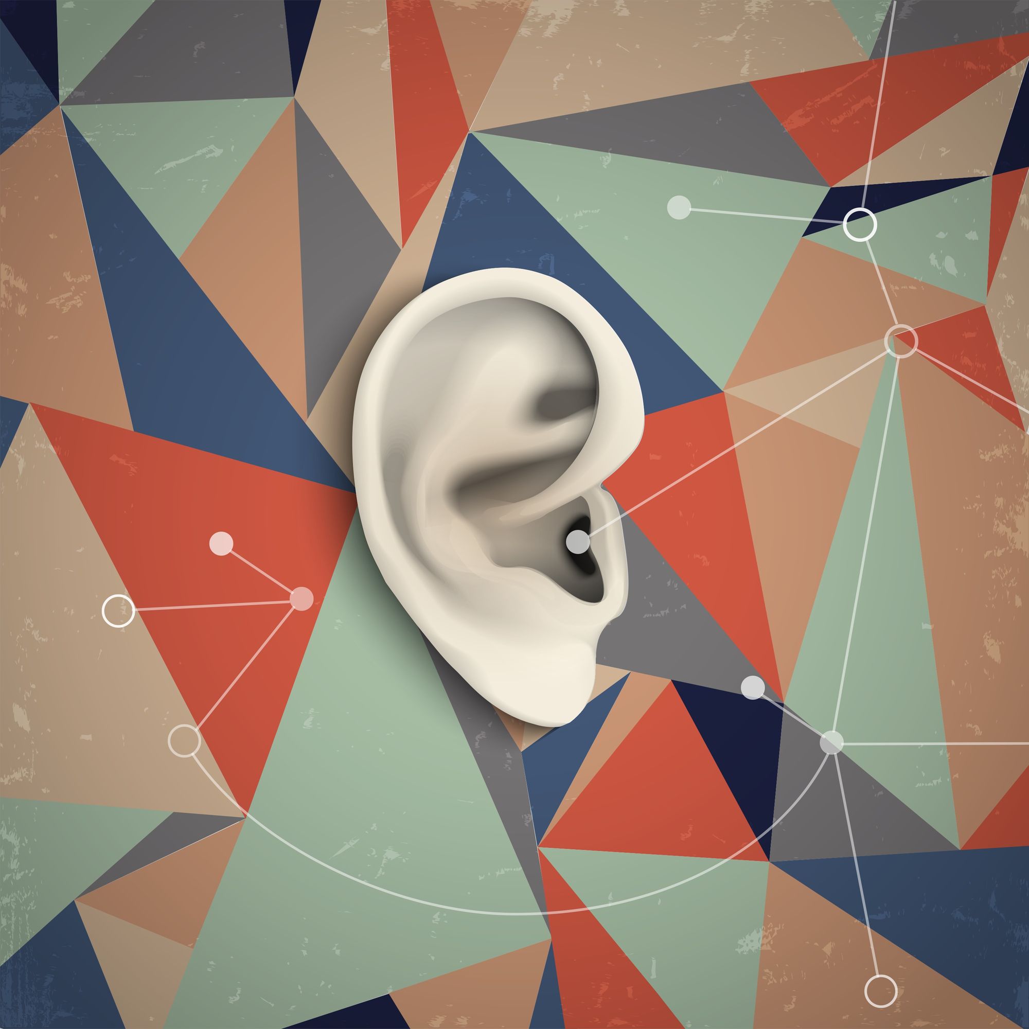
Next test the 8th Cranial Nerve. Test the cochlear component by using the Rinne and Weber's test. The vestibular component can be tested at the end of the exam by assessing balance.
Rinne Test
- Place a ringing tuning fork at the mastoid process (bone conduction) and in front of the ear (air conduction) and ask the patient which do they hear best. Air conduction should be better than bone conduction. Repeat the test on the opposite ear.
Weber’s Test
- Place a ringing tuning fork in the middle of the patient's forehead. Ask them if the sound is the same in both ears or louder on one side.
7. Cranial Nerve IX - Glossopharyngeal Nerve
Provides sensory innervation to the palate. Test the following:
Gag reflex
- Place a tongue depressor at the back of the palate and the patient should gag.
Warn the patient of the test and that it will be uncomfortable.
8. Cranial Nerve X - Vagus Nerve
Next test the Vagus Nerve which provides the motor supply to the Pharynx.
Uvula
- Note the position of the uvula. Ask the patient to say “Aaahhh”. The uvula should remain central. If it deviates, it will be towards the lesion.
9. Cranial Nerve XI - Accessory Nerve
The Accessory nerve provides motor supply to the Sternocleidomastoid and Trapezius muscles of the neck. Test them by doing the following:
Sternocleidomastoid muscle
- Test the (R) side by placing a hand on the (L) cheek and asking the patient to push their cheek against resistance. Do the opposite to test the (L) side.
Trapezius muscle
Place your hands on the patient’s shoulders and ask them to raise their shoulders against resistance.
10. Cranial Nerve XII - Hypoglossal Nerve
Test the Hypoglossal Nerve, which provides the motor supply to the tongue, by asking the patient to stick their tongue out. Observe for the following:
- Wasting (lower motor neurone lesion)
- Fasciculation - twitching movements at the surface of the tongue (lower motor neurone lesion)
- Deviation (Lower motor neurone lesion - tongue deviates towards the side of the lesion, Upper motor neurone lesion - tongue deviates away from the side of the lesion)
10. Check balance
Finish off the exam by testing the vestibular component of Cranial Nerve VIII by doing the Romberg's test.
End of examination
- Thank patient
Let the patient know you have finished examining them and thank them for their time.
2. State other exams for completion
Turn to the examiner and state what else you would do to complete the exam.
Doctor Khalid Newsletter
Join the newsletter to receive the latest updates in your inbox.
